OCT 3D Retinal Scans
Ocular Coherence Tomography: The Next Generation Eye Imaging Technology
DID YOU KNOW?
90% of vision loss can be prevented or treated*.
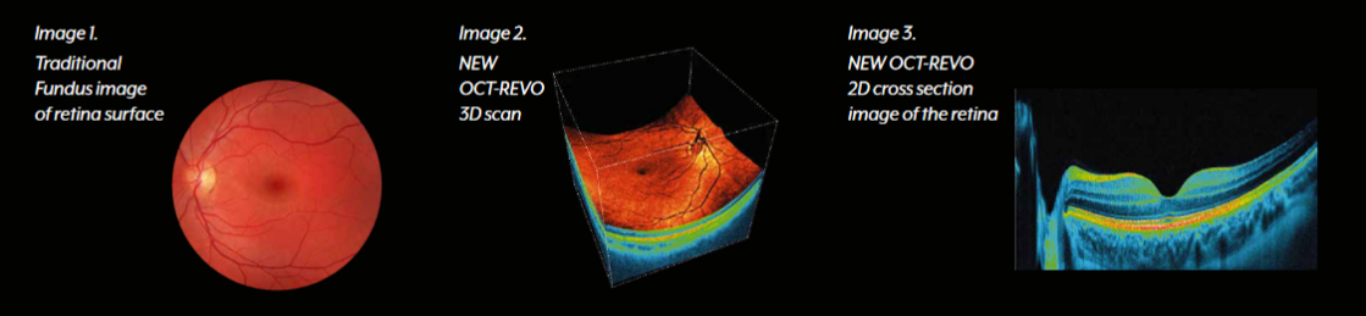
 Experience the latest technological breakthrough in eye imaging – OCT. This cutting-edge technology uses a state-of-the-art scanner to create a 3D image of the inside of your eye, similar to an optical ultrasound. Unlike traditional methods, OCT can reveal the 10 microscopic layers beneath the surface of the retina, a light-sensitive area located at the back of your eye.
The scan is quick, painless and easy to perform, taking only a few seconds to complete. It is suitable for people of all ages and can detect eye problems at an earlier stage, enabling better treatment options and improved visual outcomes.
Does the OCT scan just take a photo of the retina?
The short answer is no! This advanced technology offer a whole lot more than a picture of your retina. Below is a detailed outline of what is done and what can be detected during this scan.
A message from Parant on why the OCT is an important scan to have…

What do OCT 3D Retinal Scans show?
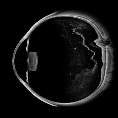
ANTERIOR CHAMBER & CORNEA SCAN
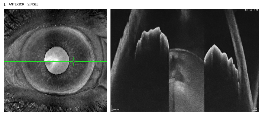
This scan shows the clarity and thickness of the cornea and confirms that the eye’s drainage channels are open and functioning.
Anterior chamber and cornea scans are most important for:
- People who have closed angle glaucoma
- People who have hyperopia (are long-sighted)
- If you have a history of eye pain &/or headaches associated with blurry vision
- People with a family history of glaucoma
- Contact lens wearers
- Anyone considering corrective laser surgery
- People who are having, or have had laser eye surgery
- People with high eye pressures (ocular hypertension)
- People of South Asian ethnicity
AXIAL LENGTH BIOMETRY SCANS
These eye scans measure the axial length of the eye, which is the distance from the front of the cornea to the retina, through the central axis of the eye. This type of eye scan is essential when monitoring progressive myopia and is considered the gold standard in myopia management.
Axial length and biometry scans are most important for:
- Myopes (short-sighted people)
- Progressive myopes (usually younger people whose myopia is still increasing)
- Those with unexplained visual problems
- People with flashes &/or floaters
- People who are having, or have had laser eye surgery
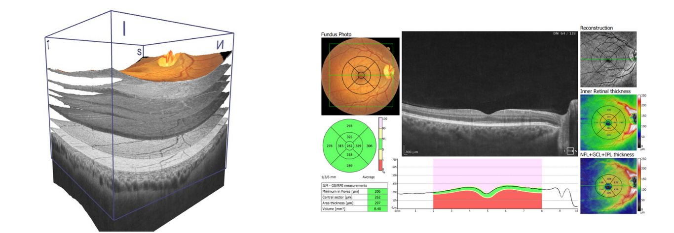
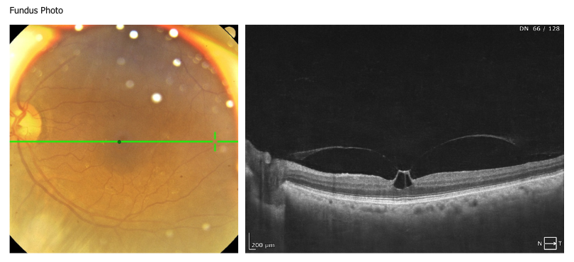
MACULA SCANS
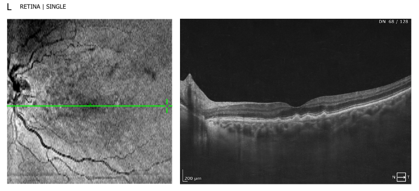
This type of eye scan shows the central area of the retina called the macula. This is the most delicate part of the eye and offers the most sensitive vision. It’s what you use to read messages on your phone, look for road signs and when you work on your computer.
The macular scans are most important if you fall into any of the categories below:
- Suffer with macular degeneration
- Have diabetes
- Have a family history of macular disease
- If you are a smoker
- If you have myopia (short-sightedness)
- For those with unexplained visual problems
- People with flashes &/or floaters
- Any unexplained loss of vision
- In cases of head or eye injuries
- Other retinal or macular disorders e.g. Epiretinal Membrane and macular hole
OPTIC NERVE SCANS
This scans shows the optic disc which is the nerve that connects the eye to the visual processing centres in the brain. The optic nerve is vital for maintaining healthy vision and its appearance can be monitored for changes as we age.
The optic disc scans are most important if you fall into any of the categories below:
- You suffer with glaucoma
- If you have a family history of glaucoma
- If you’ve been diagnosed with high eye pressures (ocular hypertension)
- Anyone who has certain neurological conditions e.g. MS
- If you have unexplained headaches
- People with unexpectedly poor vision
- Optic nerve disease e.g. optic neuritis
- Other optic nerve disorders e.g. optic disc drusen
*source: The International Agency for the Prevention of Blindness (IAPB)